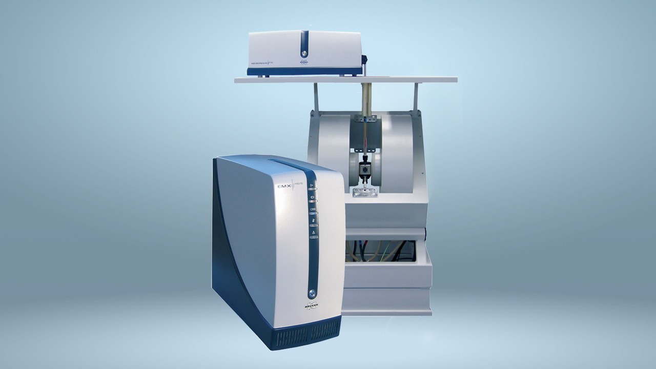
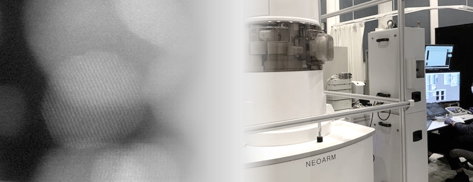
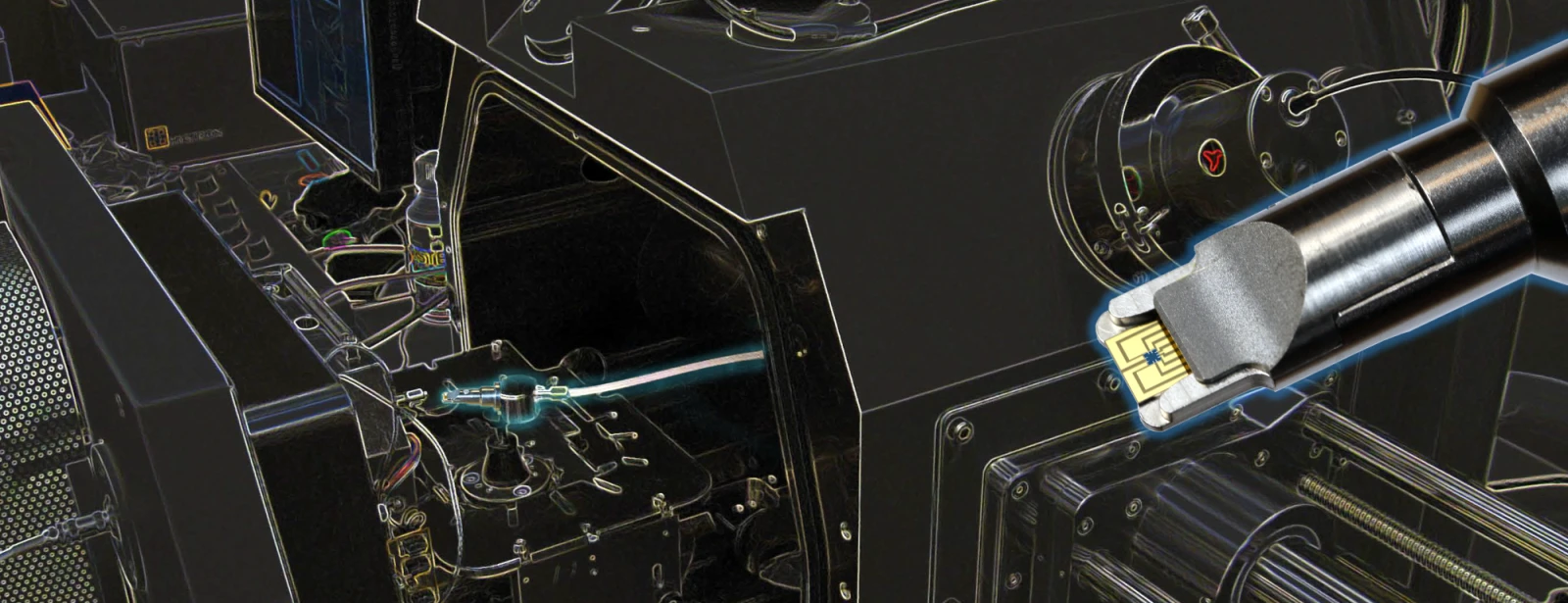
3D electron diffraction tomography (Micro-ED)
3D electron diffraction tomography approach has been developed by U.Kolb (3D diffraction tomography/instrumentation and techniques)and named asautomated diffraction tomography (ADT); 3D hkl precession data are acquired through sequentially tilting a selected nanocrystal around an arbitrary crystallographic axiswith a variable tilt step down to 1° through the maximum TEM tilting range (usually from -45° to + 45°). Such a data set contains nearly all reflections present in the covered wedge of the reciprocal space.The advantage of 3D electron diffraction tomography data collection over individual ZA collection is that data collection can begin from any arbitrary (non-oriented) section of reciprocal space without any prior knowledge of the crystal cell ;
after collection of typically 90 reciprocal space sections with single-tilt TEM holder , 3D reciprocal cell can be reconstructed automatically, crystal cell can be accurately determined (2-5% error) and 3D intensities can be measured to provide full 3D structure solution . 3D data acquisition can be done manually or automatically in some TEM platforms ; STEM Automated diffraction tomography (ADT) acquisition in nanobeam mode is especially effective for data collection from beam sensitive materials because it uses low dose illumination.
(ADT) uses software to collect diffraction patternsover a series of serial tilt increments(variable tilt step down to 1°) or in continuous tilt mode (Micro-ED)( 3D diffraction tomography/instrumentation and techniques); in this way, the generated3D(tomographic) data set of reciprocal lattice intensities can be used for structure determination. By coupling this technique with beam precession , the range and quality of the data set can be greatly improved.The combination of ADT-PED has been employed effectively to investigate complex framework structuresand beam-sensitive organic crystals., even protein crystals.( 3D diffraction tomography/proteins /organics-pharmaceuticals). Combining beam precession with dynamical refinements, it is possible to reveal Hydrogen atoms in the structure and even determine the absolute structure condiguration (3D diffraction tomography/proteins /absolute configuration .
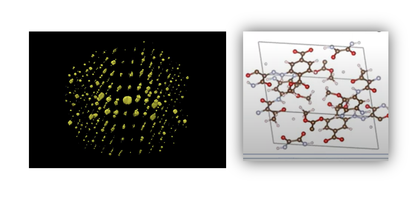
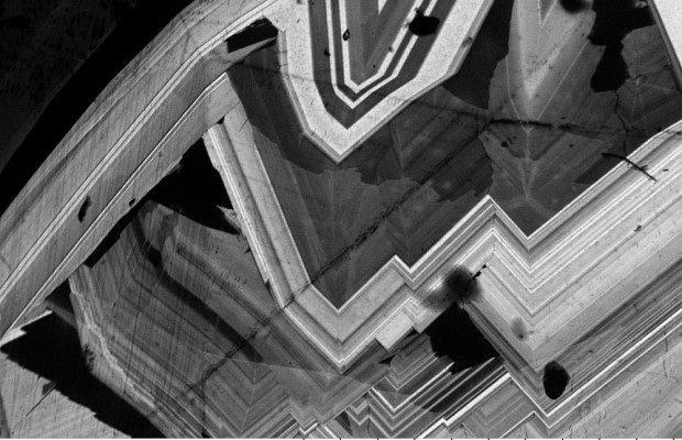
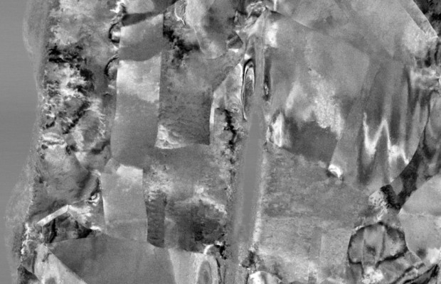
Reconstructed three dimensional reciprocal space
showing hexagonal cell (P63/mmc a= 0.396nm, c =0.536 nm) for NiTenanocystal with data resolution up to 0.8A. Data have been collected from 80 precession diffraction patterns (precession angle 17 mrad) from -40° + 40° onTecnai 30- FEG-STEM DigiSTAR (courtesyDr.E.Mugnaoili, Dr.U.Kolb Mainz Univ)


-
-
-
-

-
-
-
-
-

-
-
-
-
-
-

-

-
-
-
-
-
-
-
-

-

-

-

-

-
-
-

-
-
-
-
-
-
-
-
-
-
-
-
-
-
-
-
-
-
-
-
-
-
-

-
-
-
-
-
-
-
-
-
-
-
-
-
-
-
-
-
-
-
-

-
-
-
-
-
-

-
-
-
-
-

-
-
-

-
-

-
-
-

-
-

-
-
-
-
-
-
-
-
-
-

-
-
-

-