
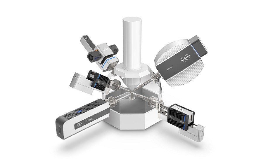
4D STEM WITH TOPSPIN PLATFORM
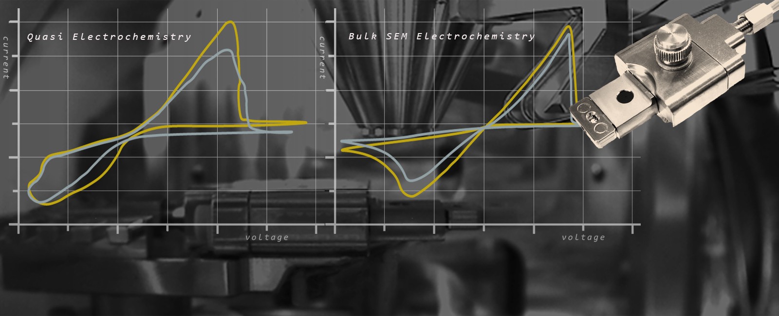
Scanned acquisition with precession electron diffraction
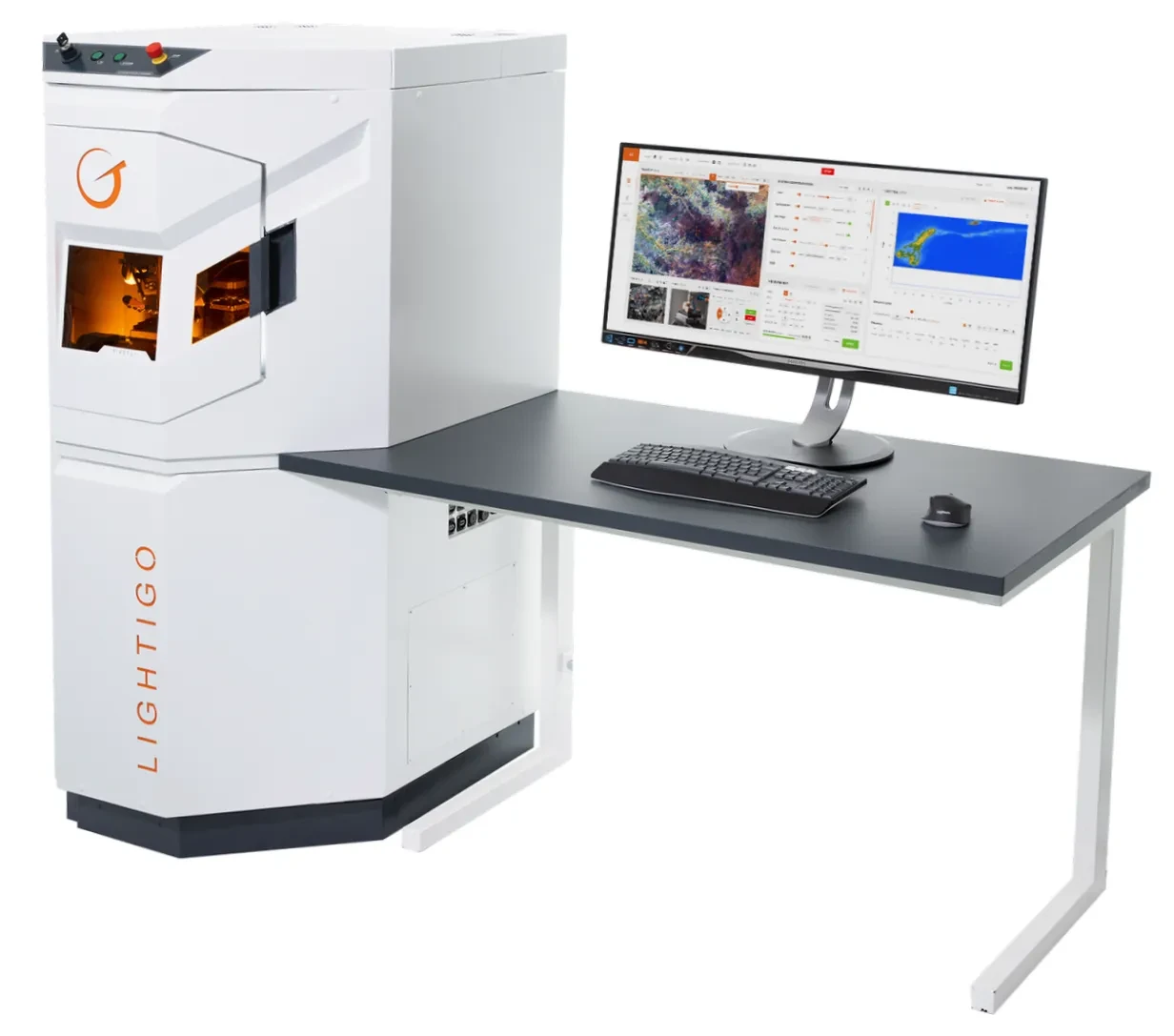
Topspin is a digital STEM, Beam Precession and Analytical Experiment Framework that offers a suite of beam precession-enabled imaging and advanced analytical experiments
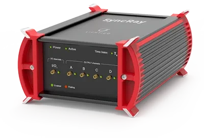
Using Digistar this system can collect precession electron diffraction (PED) patterns for each point in a scanned image.
Topspin allows researchers to routinely perform advanced analytical experiments inside an Electron Microscope in ways previously not possible.
The Topspin framework enhances TEM capabilities in several key areas:
- Digital TEM and STEM imaging
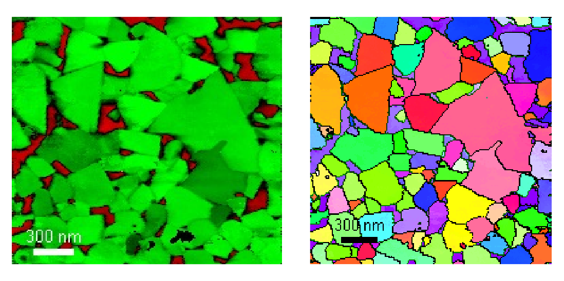
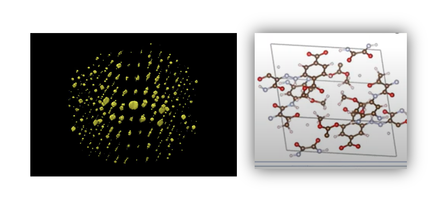
- Phase and orientation mapping (ASTAR)
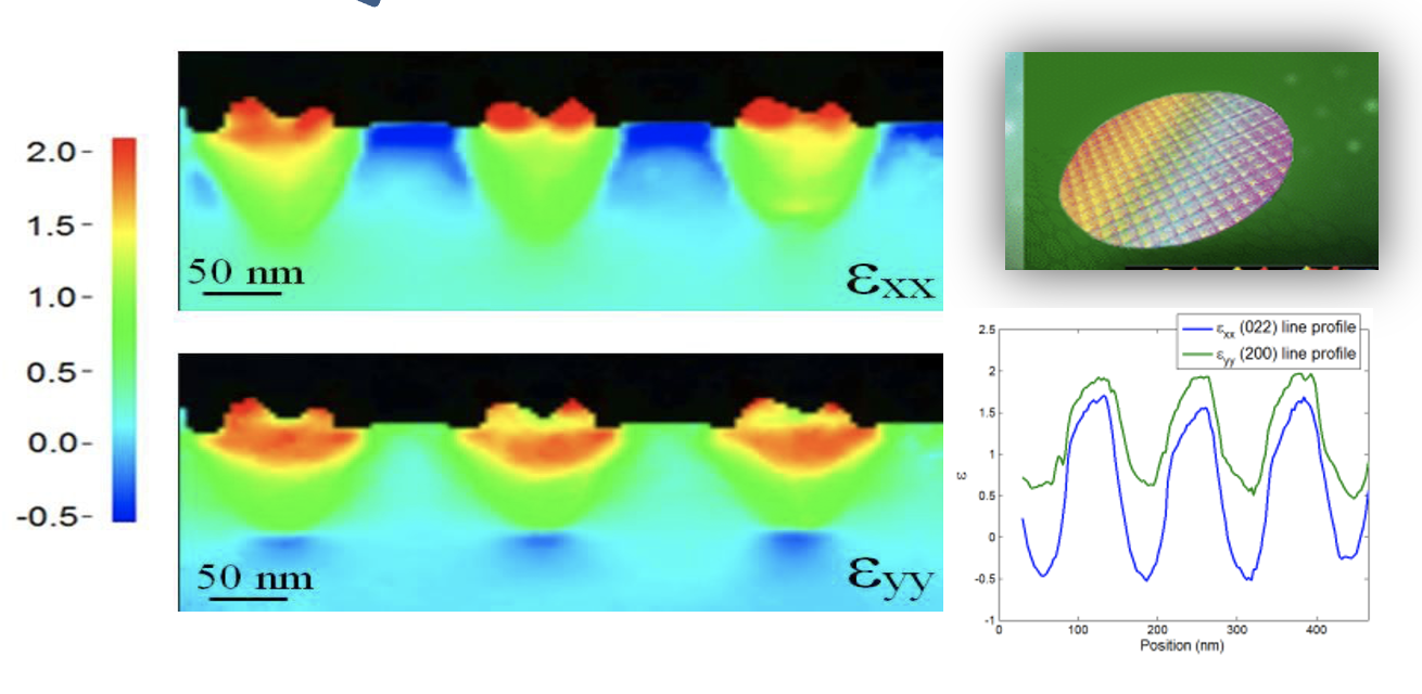
- Strain mapping analysis
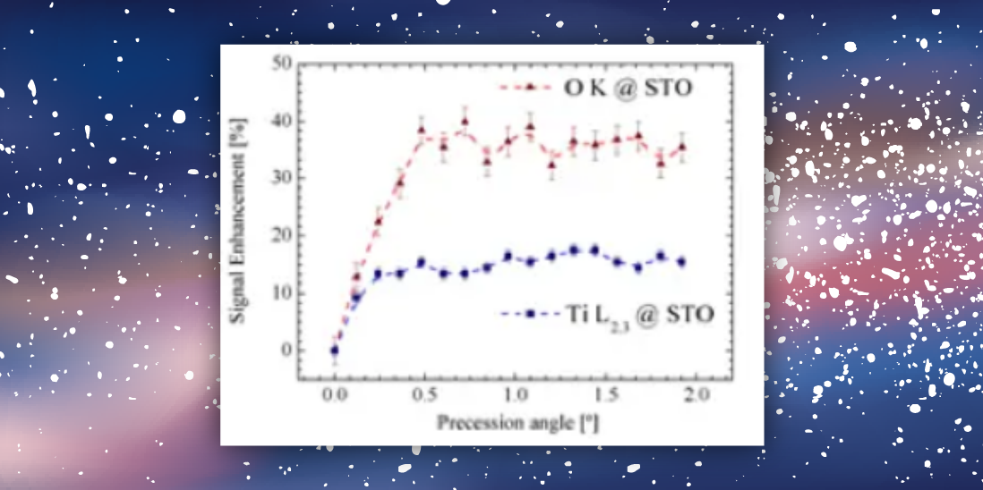
- Enhanced EDX and EELS spectroscopy
- Model-based EELS quantification

4D-STEM with Topspin PED Acquisition software*
- Synchronized beam scanning and precession with multi-signal data acquisition
- Advanced control of DigiSTAR and external optical CCD camera
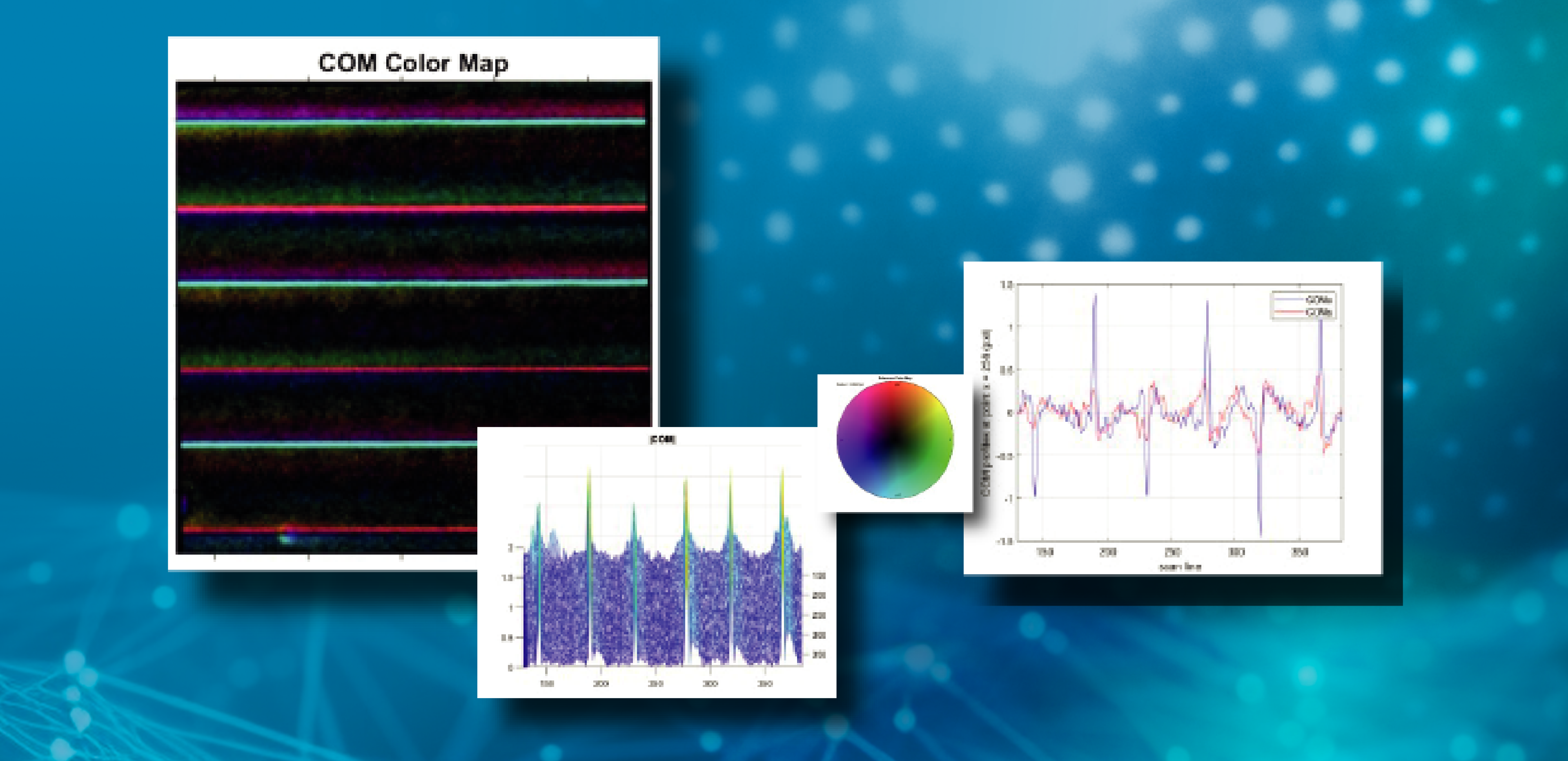
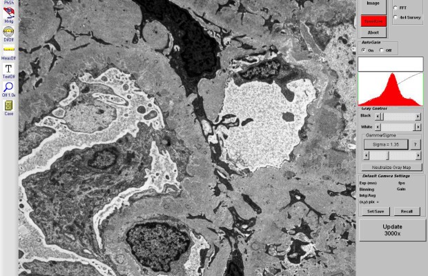
- Virtual STEM imaging with beam precession control. Virtual reference STEM image providing accurate control of scanned region. Mapping of site specific regions possible with precise positioning of region of interest, using multiple scan modes (spot, line, area)
- Virtual BF/DF via external optical CCD
- On-line distortion correction of CCD camera images
- Specific software module that allows data acquisition with high sensitivity/speed from Direct electron detectors. Data can be used with ASTAR or STRAIN.
- Drift correction available as a reference image is acquired before scanning. Drift correction window is tracked during acquisition
- Calibration of rotation between virtual STEM images and acquired diffraction patterns is automatically exported with ASTAR *.blo files
- Full 14-bit dynamic range of acquired diffraction patterns is retained in database files. Intensity scaling (brightness/contrast/gamma) for *.blo files can be readjusted after acquisition
* needs DigiSTAR
AUTOMATED BEAM COMPENSATION
- Completely automated simultaneous adjustment of 64 compensation points for best possible spatial resolution. Additional compensation points can be added as needed
- 16 bit images used for alignment
- Sub-pixel alignment accuracy at 580×580 camera resolution performed by looking at the image of the probe in order to provide correct pivot point adjustment, even at large precession angles
Diğer Ürünler
-
QUANTAX EDS for SEM -
QUANTAX WDS -
EBSD -
Micro-XRF -
Fourier 80 -
ELEXSYS -
EMXplus -
EMXmicro -
Magnettech ESR5000 -
AvanceCore -
Avance NMR -
GHz Class NMR -
ASTAR -
TEM STRAIN MAPPING ANALYSIS -
3D PRECESSION DIFFRACTION TOMOGRAPHY – MICRO ED -
e-PDF software suite -
4D-Scanning Precession Electron Diffraction (4D-SPED) -
ENHANCED EELS & EDX SPECTROSCOPY -
FireFly -
SyncRay -
3-axes Motorized Stage -
Custom designed systems -
MINISPEC MQ SERIES Contrast Agent Analyzer -
MINISPEC MQ SERIES Toothpaste Analyzer -
MINISPEC MQ SERIES Polymer Research Analyzer -
MINISPEC MQ ONE SERIES Dip on Fibers -
MINISPEC MQ ONE SERIES Hydrogen Analyzer -
MINISPEC MQ ONE SERIES Spin-Finish -
MINISPEC LF Series -
MINISPEC MQ SERIES SFC Analyzer (field-upgradeable) -
MINISPEC MQ 20 SERIESTotal Fat and Moisture -
MINISPEC MQ 20 SERIES Droplet Size Analyzer 2.0 -
MINISPEC MQ ONE SERIES Seeds Analyzer -
5 solutions in one chamber -
LUXOR Gold Coater -
LUXOR Platinum Coating -
Accessories -
SEM Backscattered Electron Detector -
SEM BSE Diode repair & supply -
Centaurus Cathodoluminescence SEM CL, BSE, YAG Detecto -
BF DF HAADF SEM STEM Detector -
HAADF Annular SEM STEM Detector -
AMT sCMOS & CCD TEM Camera Systems -
SEM EBIC Amplifier (Specimen Current Detector) & specimen holders -
Model 1064 ChipMill -
Model 1040 NanoMill® TEM specimen preparation system -
Model 1080 PicoMill® TEM specimen preparation system -
Model 1061 SEM Mill -
Model 1051 TEM Mill -
Model 1062 TrionMill -
Model 1070 NanoClean -
Model 1020 Plasma Cleaner -
Model 9030 Turbo Pumping Station -
Model 9020 Vacuum Pumping Station -
Model 9010 Vacuum Storage Container -
Model 120 Automatic Power Control -
Model 110 Automatic Twin-Jet Electropolisher -
Model 140 Digital Power Control -
Model 200 Dimpling Grinder -
Model 220 Low Temp Container -
Model 160 Specimen Grinder -
Model 2020 Advanced Tomography Holder -
Model 2021 Analytical Tomography Holder -
Model 2023 Analytical Tomography Holder for Ultra-X -
Model 2552 Cryo Cartridge Specimen Holder -
Model 2550 Cryo Transfer Tomography Holder -
Model 2040Dual-Axis Tomography Holder -
Model 2045Motorized Dual-Axis Tomography Holder -
Model 2050On-Axis Rotation Tomography Holder -
Model 2030Ultra-Narrow Gap Tomography Holder -
Model 2560Vacuum Transfer Tomography Holder -
1490 series Bulk Liquid Electrochemistry -
1405 series Optical Bulk Liquid Electrochemistry -
Liquid Flow -
Multichannel Gas Environment Heating -
MEMS Heating + Biasing -
Cryo Biasing -
Biasing Manipulator -
Tomography -
Magnetizing -
Liquid Flow -
In-Situ SEM Bulk Liquid Electrochemistry 1470 Series SEM -
SEM Gas + Heating 1300 Series – SEM -
SEM MEMS Heating + Biasing 1590 Series – SEM -
300N & 2kN vertical 3&4 point bending stage for Raman -
200N compression & horizontal bending stage -
Dual leadscrew insitu tensile stage for XRD, Optical & Synchrotron -
2kN & 5kN Tensile compression and horizontal bending stage -
200N compression & horizontal bending stage for SEM -
2kN & 5kN Tensile compression and horizontal bending stage for SEM -
300N & 2kN vertical 3&4 point bending stage for SEM -
NEW EBSD testing stages -
Dual screw tensile stage for XRD, Optical & Synchrotron -
2kN & 5kN Tensile compression and horizontal bending stage -
200N compression & horizontal bending stage -
CT500 500N in-situ tensile stage for µXCT applications -
CT5000 5kN in-situ loadcell tensile stage for X-Ray CT applications -
CT Heating and Cooling cell -20°C to +160°C for µXCT applications -
CT20K, Full Open Frame, Tensile, Compression & Torsion rig, designed for Synchrotron and room based X-ray CT imaging systems -
Biaxial & Quadaxial -
Forensic Comparison Stage for SEM -
miniFly -
Software: ESPRIT Family -
QUANTAX EDS for TEM